>>> Research: Skeletal Anthropometry
Modeling Skeletal Components from CT Data
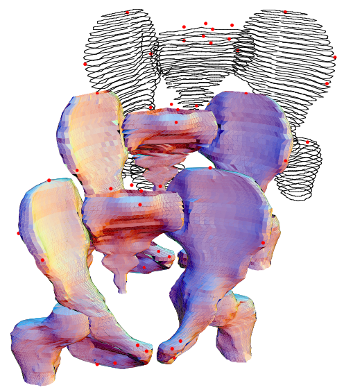
My colleagues and I have applied anthropometry methods developed for external data to the modeling of skeletal structures for application to human modeling and crash dummy design. Most finite-element models of the human body used for crash simulation are based on a single specimen, usually the Visible Human. More useful simulations could be conducted using parametric models of body structures that captured the variation across individuals. Similarly, crash dummy pelves have typically been constructed by scaling X-ray data from a few individuals, or using manual measurements from skeletal pelves. The pelves of the Hybrid-III 6YO and 10YO dummies are based on scaling of the adult Hybrid-III pelvis to match pelvis-width data from standard anthropometric surveys. Because the geometry of the pelvis is an important component of overall dummy biofidelity, we have developed a new model of the child pelvis based on CT reconstructions. We used ImageJ software from NIH (kudos to Wayne Rasband for a fantastic program), OsiriX (another fantastic open source program for processing medical imaging data), and custom software I wrote in Mathematica to generate detailed surface meshes for over 100 child pelves (ages 4 to 12). A statistical analysis based on principal components was used to generate "average" pelves for the 6YO and 10YO crash dummies.
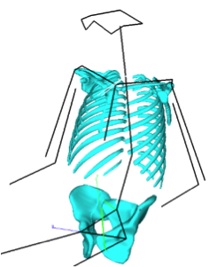
We have extended these methods to the child ribcage, clavicle, scapula, and spine. The figure to the right shows the results of predicting child skeletal geometry, surface landmarks, and joints, for a typical U.S. 10-year-old child These analyses are valuable for specifying targets for crash dummy design as well as for assessing restraint system layout. The models are able to represent not only the typical or average values, but also the variability across the population.
For more anthropometry research, see:
- Anthropometry in Automotive Postures for Drivers and Passengers
- Child Anthropometry
- Whole-Body Surface Shape Modeling
©2024 Matthew P. Reed and The University of Michigan